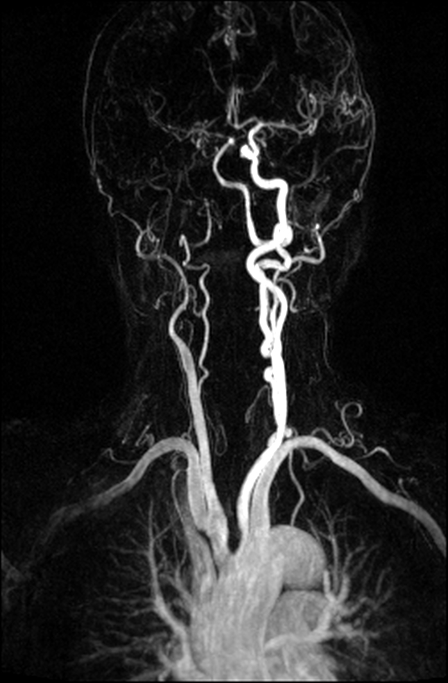

Within the past 15 years, 1.5 Tesla magnetic resonance angiography (MRA) has evolved to become an excellent non-invasive diagnostic alternative to DSA, yielding sensitivity rates of 79–97% for the detection of small UIA –. Nevertheless, due to the application of ionizing radiation and iodinated contrast agent as well as the general risk affiliated to invasive interventional procedures, DSA is associated with a 0.2%–0.5% risk for severe permanent neurological complications. ĭigital subtraction angiography (DSA) is considered the gold standard for detection of UIA. Size and shape of unruptured intracranial aneurysms (UIA) are known to be significantly affiliated with rupture rates, hence, high-quality assessment of UIA and its related features displays an important role on potential aneurysm treatment –. Rupture of intracranial aneurysm is associated with high morbidity and mortality rates, as it is known to be accountable for 80% of all subarachnaoid hemorrhages (SAH), causing 25% of all cerebrovascular-related deaths.

The funders had no role in study design, data collection and analysis, decision to publish, or preparation of the manuscript.Ĭompeting interests: The authors have declared that no competing interests exist.

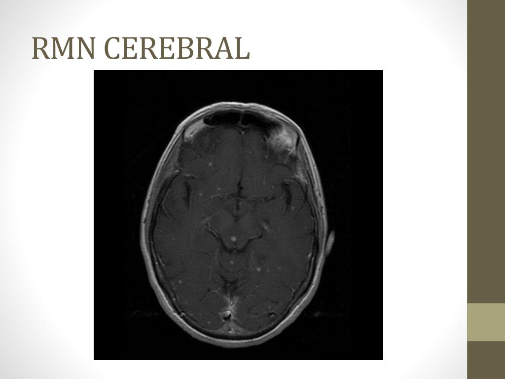
This is an open-access article distributed under the terms of the Creative Commons Attribution License, which permits unrestricted use, distribution, and reproduction in any medium, provided the original author and source are credited.įunding: An IFORES grant to KHW from the University Duisburg-Essen supported the research. Received: AugAccepted: NovemPublished: January 6, 2014Ĭopyright: © 2014 Wrede et al. PLoS ONE 9(1):Įditor: Zhuoli Zhang, Northwestern University Feinberg School of Medicine, United States of America (2014) Non-Enhanced MR Imaging of Cerebral Aneurysms: 7 Tesla versus 1.5 Tesla. Year: 2019 Type of Publication: In Proceedings Keywords: cerebral RMN hit-or-miss skeletonization isolated foreground pixel stroke Volume: 19 SGEM Book title: 19th International Multidisciplinary Scientific GeoConference SGEM 2019 Book number: 6.3 SGEM Series: International Multidisciplinary Scientific GeoConference-SGEM Pages: 229-236 Publisher address: 51 Alexander Malinov blvd, Sofia, 1712, Bulgaria SGEM supporters: Bulgarian Acad Sci Acad Sci Czech Republ Latvian Acad Sci Polish Acad Sci Russian Acad Sci Serbian Acad Sci & Arts Slovak Acad Sci Natl Acad Sci Ukraine Natl Acad Sci Armenia Sci Council Japan World Acad Sci European Acad Sci, Arts & Letters Ac Period: 9 - 11 December, 2019 ISBN: 97-99-7 ISSN: 1314-2704 Conference: 19th International Multidisciplinary Scientific GeoConference SGEM 2019, 9 - 11 December, 2019 DOI: 10.5593/sgem2019V/6.3/S08.Citation: Wrede KH, Dammann P, Mönninghoff C, Johst S, Maderwald S, Sandalcioglu IE, et al. The higher numbers of isolated foreground pixel determined using hit-or-miss transform indicate this method as a promising approach for a simple and quick evaluation of stroke. The mean ? SD values of isolated foreground pixels for stroke patients systematically exceed the corresponding values for healthy patients. We have tested the proposed algorithms using two cerebral image datasets (healthy and acute stroke patients). pixels satisfying a neighborhood configuration that corresponds to an isolated foreground pixel) were extracted using hit-or-miss and skeleton transformations. Brain MRI image was divided into eight equal sectors. This study uses the isolated foreground pixels in MRI images as a feature able to discern a stroke patient by a healthy one. Computer aided diagnosis CAD is a valuable tool to improve the ability of an average radiologist to diagnose the subtle lesions. USING GREY-SCALE HIT-OR-MISS TRANSFORM FOR DETECTION OF ISOLATED FOREGROUND PIXELS IN CEREBRAL RMN DATASETSĪbstract: MRI images contain a lot of subtle information related to various lesions which are difficult to be picked up by radiologists.


 0 kommentar(er)
